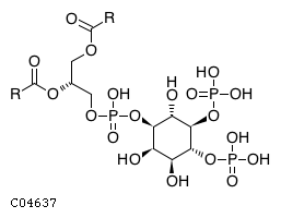
CHEBI:18348
| Name | 1-phosphatidyl-1D-myo-inositol 4,5-bisphosphate |  Download: mol | sdf |
| Synonyms | 01,2-diacyl-sn-glycero-3-phospho-(1'-myo-inositol-4',5'-bisphosphate); 1-phosphatidyl-1d-myo-inositol 4,5-bisphosphate; 1-phosphatidyl-d-myo-inositol 4,5-bisphosphate; 2,3-bis(alkanoyloxy)propyl (1r,2r,3s,4r,5r,6s)-2,3,6-trihydroxy-4,5-bis(phosphonatooxy)cyclohexyl phosphate; Phosphatidyl-myo-inositol 4,5-bisphosphate; Phosphatidylinositol-4,5-bisphosphate; Pi(4,5)p2; Pip2; Pipp; Pips; Ptdins(4,5)p2; Ptsins(4,5)p2; Ptsins-4,5-p2; | |
| Definition | A phosphatidylinositol bisphosphate in which the two phosphate groups are at C-4 and C-5 of the inositol moiety which has the 1D-myo configuration. | |
| Molecular Weight (Exact mass) | NA | |
| Molecular Formula | C11H19O19P3R2 | |
| SMILES | [H][C@@](COC([*])=O)(COP(O)(=O)O[C@@H]1[C@H](O)[C@H](O)[C@@H](OP(O)(O)=O)[C@H](OP(O)(O)=O)[C@H]1O)OC([*])=O | |
| InChI | InChI=1S/C6H17O21P5/c7-1-2(23-28(8,9)10)4(25-30(14,15)16)6(27-32(20,21)22)5(26-31(17,18)19)3(1)24-29(11,12)13/h1-7H,(H2,8,9,10)(H2,11,12,13)(H2,14,15,16)(H2,17,18,19)(H2,20,21,22)/t1-,2-,3+,4-,5-,6-/m0/s1 | |
| InChI Key | CTPQAXVNYGZUAJ-PTQMNWPWSA-N | |
| Crosslinking annotations | KEGG:C04637 | ChEBI:18348 | PubChem:7226 | |
| Pathway ID | Pathway Name | Pathway Description (KEGG) |
| map00562 | Inositol phosphate metabolism | NA |
| map01100 | Metabolic pathways | NA |
| map04070 | Phosphatidylinositol signaling system | NA |
| map04072 | Phospholipase D signaling pathway | Phospholipase D (PLD) is an essential enzyme responsible for the production of the lipid second messenger phosphatidic acid (PA), which is involved in fundamental cellular processes, including membrane trafficking, actin cytoskeleton remodeling, cell proliferation and cell survival. PLD activity can be stimulated by a large number of cell surface receptors and is elaborately regulated by intracellular factors, including protein kinase C isoforms, small GTPases of the ARF, Rho and Ras families and the phosphoinositide, phosphatidylinositol 4,5-bisphosphate (PIP2). The PLD-produced PA activates signaling proteins and acts as a node within the membrane to which signaling proteins translocate. Several signaling proteins, including Raf-1 and mTOR, directly bind PA to mediate translocation or activation, respectively. |
| map04144 | Endocytosis | Endocytosis is a mechanism for cells to remove ligands, nutrients, and plasma membrane (PM) proteins, and lipids from the cell surface, bringing them into the cell interior. Transmembrane proteins entering through clathrin-dependent endocytosis (CDE) have sequences in their cytoplasmic domains that bind to the APs (adaptor-related protein complexes) and enable their rapid removal from the PM. In addition to APs and clathrin, there are numerous accessory proteins including dynamin. Depending on the various proteins that enter the endosome membrane, these cargoes are sorted to distinct destinations. Some cargoes, such as nutrient receptors, are recycled back to the PM. Ubiquitylated membrane proteins, such as activated growth-factor receptors, are sorted into intraluminal vesicles and eventually end up in the lysosome lumen via multivesicular endosomes (MVEs). There are distinct mechanisms of clathrin-independent endocytosis (CIE) depending upon the cargo and the cell type. |
| map04151 | PI3K-Akt signaling pathway | The phosphatidylinositol 3' -kinase(PI3K)-Akt signaling pathway is activated by many types of cellular stimuli or toxic insults and regulates fundamental cellular functions such as transcription, translation, proliferation, growth, and survival. The binding of growth factors to their receptor tyrosine kinase (RTK) or G protein-coupled receptors (GPCR) stimulates class Ia and Ib PI3K isoforms, respectively. PI3K catalyzes the production of phosphatidylinositol-3,4,5-triphosphate (PIP3) at the cell membrane. PIP3 in turn serves as a second messenger that helps to activate Akt. Once active, Akt can control key cellular processes by phosphorylating substrates involved in apoptosis, protein synthesis, metabolism, and cell cycle. |
| map04510 | Focal adhesion | Cell-matrix adhesions play essential roles in important biological processes including cell motility, cell proliferation, cell differentiation, regulation of gene expression and cell survival. At the cell-extracellular matrix contact points, specialized structures are formed and termed focal adhesions, where bundles of actin filaments are anchored to transmembrane receptors of the integrin family through a multi-molecular complex of junctional plaque proteins. Some of the constituents of focal adhesions participate in the structural link between membrane receptors and the actin cytoskeleton, while others are signalling molecules, including different protein kinases and phosphatases, their substrates, and various adapter proteins. Integrin signaling is dependent upon the non-receptor tyrosine kinase activities of the FAK and src proteins as well as the adaptor protein functions of FAK, src and Shc to initiate downstream signaling events. These signalling events culminate in reorganization of the actin cytoskeleton; a prerequisite for changes in cell shape and motility, and gene expression. Similar morphological alterations and modulation of gene expression are initiated by the binding of growth factors to their respective receptors, emphasizing the considerable crosstalk between adhesion- and growth factor-mediated signalling. |
| map04666 | Fc gamma R-mediated phagocytosis | Phagocytosis plays an essential role in host-defense mechanisms through the uptake and destruction of infectious pathogens. Specialized cell types including macrophages, neutrophils, and monocytes take part in this process in higher organisms. After opsonization with antibodies (IgG), foreign extracellular materials are recognized by Fc gamma receptors. Cross-linking of Fc gamma receptors initiates a variety of signals mediated by tyrosine phosphorylation of multiple proteins, which lead through the actin cytoskeleton rearrangements and membrane remodeling to the formation of phagosomes. Nascent phagosomes undergo a process of maturation that involves fusion with lysosomes. The acquisition of lysosomal proteases and release of reactive oxygen species are crucial for digestion of engulfed materials in phagosomes. |
| map04670 | Leukocyte transendothelial migration | Leukocyte migaration from the blood into tissues is vital for immune surveillance and inflammation. During this diapedesis of leukocytes, the leukocytes bind to endothelial cell adhesion molecules (CAM) and then migrate across the vascular endothelium. A leukocyte adherent to CAMs on the endothelial cells moves forward by leading-edge protrusion and retraction of its tail. In this process, alphaL /beta2 integrin activates through Vav1, RhoA, which subsequently activates the kinase p160ROCK. ROCK activation leads to MLC phosphorylation, resulting in retraction of the actin cytoskeleton. Moreover, Leukocytes activate endothelial cell signals that stimulate endothelial cell retraction during localized dissociation of the endothelial cell junctions. ICAM-1-mediated signals activate an endothelial cell calcium flux and PKC, which are required for ICAM-1 dependent leukocyte migration. VCAM-1 is involved in the opening of the endothelial passage through which leukocytes can extravasate. In this regard, VCAM-1 ligation induces NADPH oxidase activation and the production of reactive oxygen species (ROS) in a Rac-mediated manner, with subsequent activation of matrix metallopoteinases and loss of VE-cadherin-mediated adhesion. |
| map04725 | Cholinergic synapse | Acetylcholine (ACh) is a neurotransmitter widely distributed in the central (and also peripheral, autonomic and enteric) nervous system (CNS). In the CNS, ACh facilitates many functions, such as learning, memory, attention and motor control. When released in the synaptic cleft, ACh binds to two distinct types of receptors: Ionotropic nicotinic acetylcholine receptors (nAChR) and metabotropic muscarinic acetylcholine receptors (mAChRs). The activation of nAChR by ACh leads to the rapid influx of Na+ and Ca2+ and subsequent cellular depolarization. Activation of mAChRs is relatively slow (milliseconds to seconds) and, depending on the subtypes present (M1-M5), they directly alter cellular homeostasis of phospholipase C, inositol trisphosphate, cAMP, and free calcium. In the cleft, ACh may also be hydrolyzed by acetylcholinesterase (AChE) into choline and acetate. The choline derived from ACh hydrolysis is recovered by a presynaptic high-affinity choline transporter (CHT). |
| map04810 | Regulation of actin cytoskeleton | NA |
| map05131 | Shigellosis | Shigellosis, or bacillary dysentery, is an intestinal infection caused by Shigella, a genus of enterobacteria. Shigella are potential food-borne pathogens that are capable of colonizing the intestinal epithelium by exploiting epithelial-cell functions and circumventing the host innate immune response. During basolateral entry into the host-cell cytoplasm, Shigella deliver a subset of effectors into the host cells through the type III secretion system. The effectors induce membrane ruffling through the stimulation of the Rac1-WAVE-Arp2/3 pathway, enabling bacterial entry into the epithelial cells. During multiplication within the cells, Shigella secrete another subset of effectors. VirG induces actin polymerization at one pole of the bacteria, allowing the bacteria to spread intracellularly and to infect adjacent cells. OspF, OspG and IpaH(9.8) downregulate the production of proinflammatory cytokines such as IL-8, helping bacteria circumvent the innate immune response. |
| map05132 | Salmonella infection | Salmonella infection usually presents as a self-limiting gastroenteritis or the more severe typhoid fever and bacteremia. The common disease-causing Salmonella species in human is a single species, Salmonella enterica, which has numerous serovars.Following intestinal colonization Salmonella inject effector proteins into the host cells using a type III secretion system (T3SS), T3SS1. Then a small group of effector proteins induce rearrangement of the actin cytoskeleton resulting in membrane ruffles and rapid internalization of the bacteria.The T3SS2 is responsible for translocating effector proteins that direct Salmonella-containing vacuole (SCV) maturation. The majority of the bacteria are known to survive and replicate in SCV. |

